

 Opinion: What does redemption really mean?
 A look ahead at the ‘preferred design’ for Concord’s new police headquarters
 Concord may finally buy long-closed rail line with hopes of creating city-spanning trail
 ZBA appointment to be reconsidered at Monday Council meeting
 Opinion: As people of faith, the state budget offends our values
 Summer baseball peaks as Concord faces Weare in American Legion play
 Layoffs, grant funding cuts take effect at New Hampshire’s Council on the Arts
 Public universities scrub DEI webpages in response to state budget prohibitions
 ‘Time for me to move on’ — Heartsong Healing Center closes in Hooksett as owner retires

New Cheers owners honor restaurant’s original menu while building something fresh
Greg Tandy started asking around when he became the new owner of Cheers.

‘Love you more’: Concord community remembers Glenn Chrzan
Deborah Eckland stood in front of section 47 at the New Hampshire State Veterans Cemetery, facing a small crowd of about a dozen people. She wiped her eyes with a white handkerchief.
Most Read
 New Hampshire targets sexual exploitation and human trafficking inside massage parlors
 New Hampshire legalizes public alcohol consumption in designated ‘social districts’
 State rules Epsom must pay open-enrollment tuition to other school districts, despite its refraining from the program
 ‘A little piece of everything I like’: New Pittsfield barbershop brings more than a haircut to downtown
 Remembered: Friends recall stories about the lives of those who died without housing
 Town turmoil: Chichester town administrator resigns again
Editors Picks
 A Webster property was sold for unpaid taxes in 2021. Now, the former owner wants his money back
 Report to Readers: Your support helps us produce impactful reporting
 City prepares to clear, clean longstanding encampments in Healy Park
 Productive or poisonous? Yearslong clubhouse fight ends with council approval

Sports

Youngsters Richardson and Hakala move on, and veterans crash out at 122nd State Amateur Championship
ROCHESTER — Every golfer has A routine. Superstitions, lucky charms and a specific way of finding a mental sweet spot aren’t specific to the sport.
 Athlete of the Week: Grace Saysaw, Concord High School
 Local golfers tee off at 122nd Amateur Championship
 As Concord’s Gavin Richardson places second at golf Junior Amateur, young players look ahead to the 122nd State Amateur Championship
 Sunapee’s Bryce Whitlow keeps memory of above-average MLB players alive through social media page ‘MLB Hall of (Pretty) Good’
Opinion

Opinion: Trumpism in a dying democracy
 Opinion: What Coolidge’s century-old decision can teach us today
 Opinion: The art of diplomacy
 Opinion: After Roe: Three years of resistance, care and community
 Opinion: Iran and Gaza: A U.S. foreign policy of barbarism

Your Daily Puzzles
An approachable redesign to a classic. Explore our "hints."
A quick daily flip. Finally, someone cracked the code on digital jigsaw puzzles.
Chess but with chaos: Every day is a unique, wacky board.
Word search but as a strategy game. Clearing the board feels really good.
Align the letters in just the right way to spell a word. And then more words.
Politics

New Hampshire school phone ban could be among strictest in the country
When Gov. Kelly Ayotte called on the state legislature to pass a school phone ban in January, the pivotal question wasn’t whether the widely popular policy would pass but how far it would go.
 Concord became a Housing Champion. Now, state lawmakers could eliminate the funding.
 ‘A wild accusation’: House votes to nix Child Advocate after Rep. suggests legislative interference
  Sununu decides he won’t run for Senate despite praise from Trump
 Town elections offer preview of citizenship voting rules being considered nationwide
e-Edition
Arts & Life

Lavender haze: Purple fields bloom at Warner farm
The lavender fields at Pumpkin Blossom Farm in Warner are open for Harvest Days.
 Arts in the Park returns for July
 Hopkinton art gallery showcases “Creativity Beyond Convention”
 AROUND CONCORD: Your guide to free summer music
 Around Concord: Steps to nowhere – but it used to be somewhere
Obituaries
 Herbert Little
Concord, NH - Herbert E. "Herb" Little died on the evening of July 6, 2025, at Havenwood Heritage Heights in the room he shared with his loving wife Debby who was, as always, by his side. He was 97 years old having been born on Septembe... remainder of obit for Herbert Little
 Anna Marie O'Reilly
Contoocook, NH - Anna Marie O'Reilly, 21, of Contoocook, passed away unexpectedly at home on Friday, July 4, 2025. She was born on June 10, 2004, in Concord, NH to her parents, Joseph P. O'Reilly and Donna M. (Phillips) O'Reilly. Anna g... remainder of obit for Anna Marie O'Reilly
 Mary Ann Hermman
Baltimore, MD - Mary Ann Hermann died in her home in Baltimore on July 5, 2025, following a battle with cancer. She was 91. Mary Ann was a retired psychiatric nurse who worked in hospitals and visiting patients at their homes in Cuba, ... remainder of obit for Mary Ann Hermman
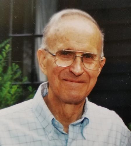
 David Franklin Whitney
Hillsboro, NH - David F. Whitney 87, a lifelong resident of Hillsboro, NH., joined his bride Patricia A. (Wing) Whitney in Heaven Thursday June 26,2025. Born September 10, 1937 to Harry L. and Harriett S. Whitney at their home on Wh... remainder of obit for David Franklin Whitney